Van Gieson Stain Pathology Outlines: A Diagnostic Tool
The van Gieson stain is a histological staining technique used in pathology to detect and visualize elastic and collagen fibers in tissue samples. This diagnostic tool has been widely used in medical research and diagnosis, providing valuable insights into the structure and composition of tissues. With its ability to highlight specific fiber types, the van Gieson stain has become an essential component of pathology outlines, helping medical professionals to identify and understand various diseases and conditions.
Understanding the Van Gieson Stain
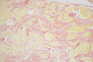
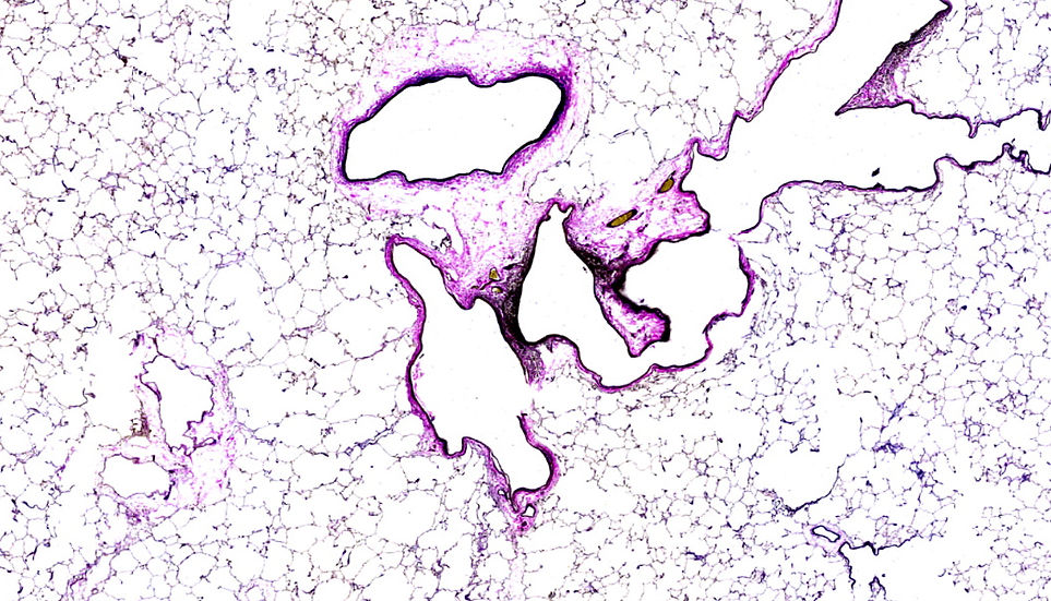
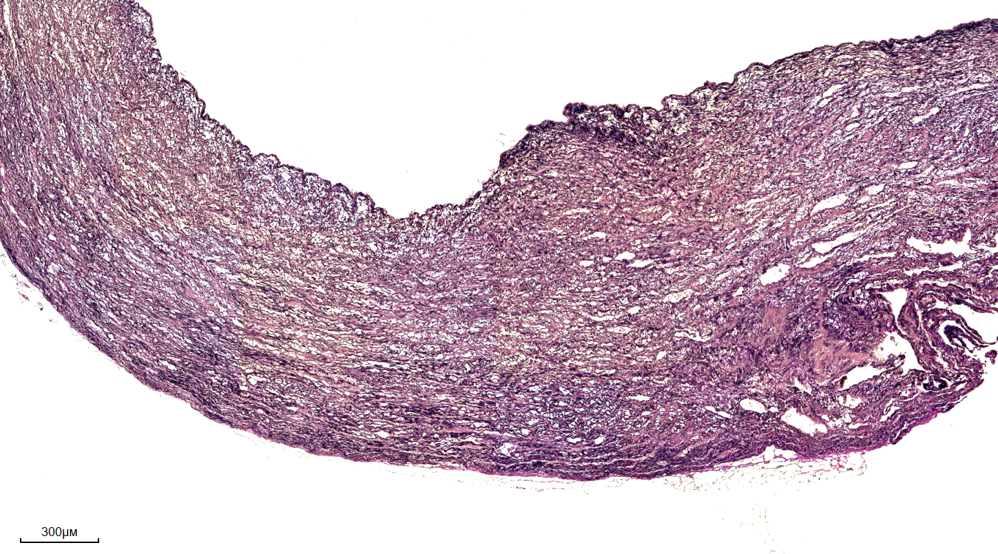
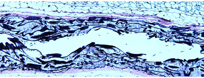
The van Gieson stain is a type of special stain that uses a combination of dyes to selectively stain elastic and collagen fibers in tissue samples. The stain works by using a specific sequence of dyes, which bind to the fibers and produce a distinctive coloration. Elastic fibers are typically stained black, while collagen fibers are stained red or pink. This coloration allows medical professionals to easily identify and distinguish between different fiber types, which is crucial for diagnosing and understanding various diseases and conditions.

Applications of the Van Gieson Stain
The van Gieson stain has a wide range of applications in pathology, including the diagnosis of diseases such as pulmonary fibrosis, atherosclerosis, and cancer. By highlighting elastic and collagen fibers, the stain allows medical professionals to assess the extent of tissue damage and identify specific patterns of fiber deposition. For example, in the case of pulmonary fibrosis, the van Gieson stain can be used to detect the accumulation of collagen fibers in the lungs, which is a characteristic feature of the disease.

Benefits and Limitations of the Van Gieson Stain
The van Gieson stain offers several benefits, including its ability to provide high-quality images of elastic and collagen fibers, its relatively simple staining protocol, and its wide range of applications in pathology. However, the stain also has some limitations, including its potential to produce variable results depending on the quality of the tissue sample and the staining protocol used. Additionally, the van Gieson stain may not be suitable for all types of tissue samples, and other staining techniques may be required to provide a comprehensive diagnosis.
Conclusion
In conclusion, the van Gieson stain is a valuable diagnostic tool in pathology, providing medical professionals with a powerful means of detecting and visualizing elastic and collagen fibers in tissue samples. With its ability to highlight specific fiber types, the van Gieson stain has become an essential component of pathology outlines, helping to advance our understanding of various diseases and conditions. While the stain has its limitations, its benefits make it a widely used and respected technique in the field of pathology.
Celnovte
Celnovte
Verhoeff Van Gieson Stain | Elastic & Collagen Fiber Detection | IHisto
Verhoeff Van Gieson Stain | Elastic & Collagen Fiber Detection | iHisto
Verhoeff-Van Gieson Stain(VVG)_規格表 - BioTnA
Verhoeff-Van Gieson stain(VVG)_規格表 - BioTnA
Verhoeff-van Gieson Stain: A Special Histology Stain For Elastic Fibers
Verhoeff-van Gieson Stain: A Special Histology Stain for Elastic Fibers